

Images
Sample images obtained by spinning-disk confocal microscopy, taken by C. Scheiermann with a similiar setup
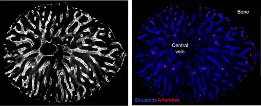
Transverse-shaved whole-mount images of the mouse femoral bone marrow stained with antibodies directed against VE-cadherin and PECAM-1 (white, left image, or blue, right image) highlighting the whole vasculature and Sca-1 (red, right image), marking arterioles. (Image by C. Scheiermann; taken from Kunisaki et al., Nature 2013)