

Images
Imaging of leukocyte intraluminal crawling, diapedesis, and interstitial chemotaxis in the mouse ear by 2-photon microscopy
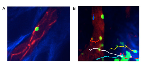
Panel A illustrates the intravascular crawling of gfp+ monocyte in the CX3CR1-gfp/+ mouse. The panel shows the positioning of crawling monocyte with respect to the endothelial junctions (red - anti-CD31 Abs).
B: Transmigration and directional interstitial migration of neutrophils (LysM-eGFP) to the steril injury site´.
Bone marrow imaging by 2-photon microscopy
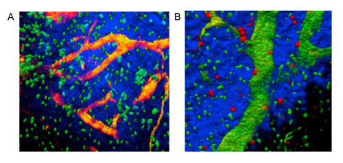
Panel A illustrates the normal micro-anatomy of the mouse calvarian bone. Blue, bone (second harmonic signal); yellowish, vasculature (FITC/TRITC-dextran); green, genetically marked osteoblasts (eGFP).
The panel B shows the positioning of HSPCs in the bone marrow compartment. HSPCs (red, CMTMR) were prospectively sorted and adoptively transferred into recipient mice, expressing eGFP under the control of an osteoblast-specific promoter.